Fossa Poplitea Anatomy: A Comprehensive Guide To The Popliteal Fossa
The fossa poplitea anatomy is a fascinating area of study that plays a crucial role in understanding the human body's functional mechanics. Located at the back of the knee, this diamond-shaped depression serves as a vital pathway for nerves, blood vessels, and lymphatics. Whether you're a student of anatomy, a healthcare professional, or simply someone interested in the intricacies of the human body, gaining insights into the fossa poplitea anatomy can deepen your appreciation for how the body operates. This article will explore its structure, function, clinical relevance, and much more, ensuring you walk away with a comprehensive understanding.
The fossa poplitea anatomy is not just a structural component of the knee; it is a hub of activity. The popliteal fossa houses essential structures such as the popliteal artery, vein, and the tibial and common fibular nerves. These components work together to facilitate movement, circulation, and sensation in the lower limbs. Understanding this anatomy can help in diagnosing and treating various conditions, such as popliteal artery aneurysms or nerve entrapments. With its intricate design, the fossa poplitea anatomy serves as a gateway to exploring the interconnectedness of the body's systems.
As we delve deeper into this topic, we will uncover the layers of complexity that make the fossa poplitea anatomy so remarkable. From its boundaries and contents to its clinical implications, this guide aims to provide a thorough overview that is both informative and engaging. Whether you're looking to enhance your knowledge or seeking practical applications, this article will serve as a valuable resource. So, let's begin our journey into the world of the popliteal fossa and discover what makes it such a vital part of human anatomy.
Read also:Tom Selleck On Treat Williams Death A Heartfelt Tribute To A Hollywood Legend
Table of Contents
- What is Fossa Poplitea Anatomy?
- Boundaries of the Popliteal Fossa
- Contents of the Popliteal Fossa
- Why is the Popliteal Fossa Important?
- Common Conditions Affecting the Popliteal Fossa
- How to Examine the Popliteal Fossa?
- Fossa Poplitea Anatomy in Clinical Practice
- Frequently Asked Questions About Fossa Poplitea Anatomy
What is Fossa Poplitea Anatomy?
The fossa poplitea anatomy refers to the anatomical structure located at the posterior aspect of the knee joint. This diamond-shaped depression is formed by the convergence of muscles, bones, and ligaments, creating a space that houses critical neurovascular structures. The popliteal fossa is a key area for both movement and circulation, making it an essential topic in the study of human anatomy.
Understanding the fossa poplitea anatomy begins with recognizing its role as a conduit for nerves and blood vessels. The popliteal artery, one of the primary blood vessels in the lower limb, runs through this area, supplying oxygenated blood to the leg and foot. Similarly, the popliteal vein and lymphatics ensure the return of deoxygenated blood and lymph to the heart. The tibial and common fibular nerves also traverse the fossa, providing motor and sensory functions to the lower leg and foot.
What sets the fossa poplitea anatomy apart is its unique position as a transition zone. It connects the thigh to the leg, allowing for the seamless transfer of forces during movement. Whether you're walking, running, or simply standing, the structures within the popliteal fossa work in harmony to support these activities. This makes it a focal point for diagnosing and treating conditions that affect mobility and circulation.
Why Study the Fossa Poplitea Anatomy?
Studying the fossa poplitea anatomy is essential for several reasons. First, it provides a foundation for understanding the interconnectedness of the body's systems. The popliteal fossa is a microcosm of how muscles, nerves, and blood vessels collaborate to enable movement and maintain homeostasis. Second, knowledge of this anatomy is critical for diagnosing and treating conditions such as popliteal artery aneurysms, nerve entrapments, and deep vein thrombosis.
Moreover, the fossa poplitea anatomy is a frequent focus in medical education and clinical practice. Surgeons, radiologists, and physiotherapists often rely on a detailed understanding of this area to perform procedures, interpret imaging, and develop rehabilitation plans. By mastering the intricacies of the popliteal fossa, healthcare professionals can improve patient outcomes and enhance their expertise.
Boundaries of the Popliteal Fossa
The fossa poplitea anatomy is defined by specific anatomical boundaries that contribute to its diamond shape. These boundaries are formed by muscles, bones, and ligaments, each playing a unique role in stabilizing and protecting the structures within the fossa.
Read also:Dorothy Jo Gideon A Comprehensive Guide To Her Life And Legacy
Superior Boundaries
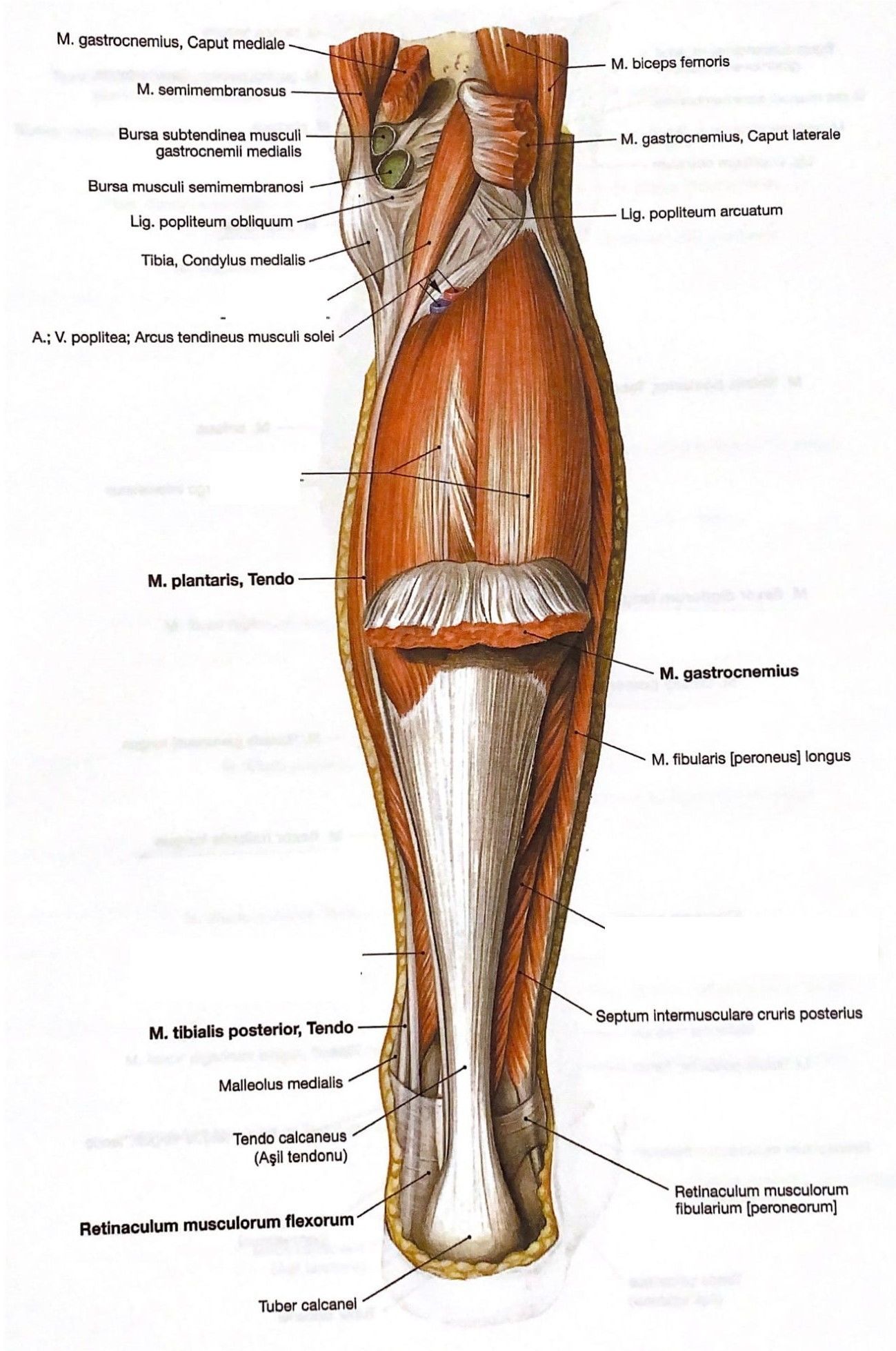
The superior boundaries of the fossa poplitea anatomy are formed by two muscles: the biceps femoris laterally and the semimembranosus and semitendinosus muscles medially. These muscles converge at the posterior aspect of the knee, creating the upper edges of the diamond-shaped depression. The biceps femoris is responsible for knee flexion and lateral rotation, while the semimembranosus and semitendinosus contribute to knee flexion and medial rotation.
Inferior Boundaries
The inferior boundaries of the fossa poplitea anatomy are defined by the lateral and medial heads of the gastrocnemius muscle. These powerful muscles are primarily involved in plantarflexion of the foot and also help stabilize the knee joint. Together, they form the lower edges of the popliteal fossa, completing its diamond shape.
Understanding these boundaries is crucial for grasping the overall structure of the fossa poplitea anatomy. The convergence of these muscles not only creates the physical space of the fossa but also provides a protective enclosure for the neurovascular structures within. This arrangement ensures that the popliteal artery, vein, and nerves are safeguarded during movement and weight-bearing activities.
Contents of the Popliteal Fossa
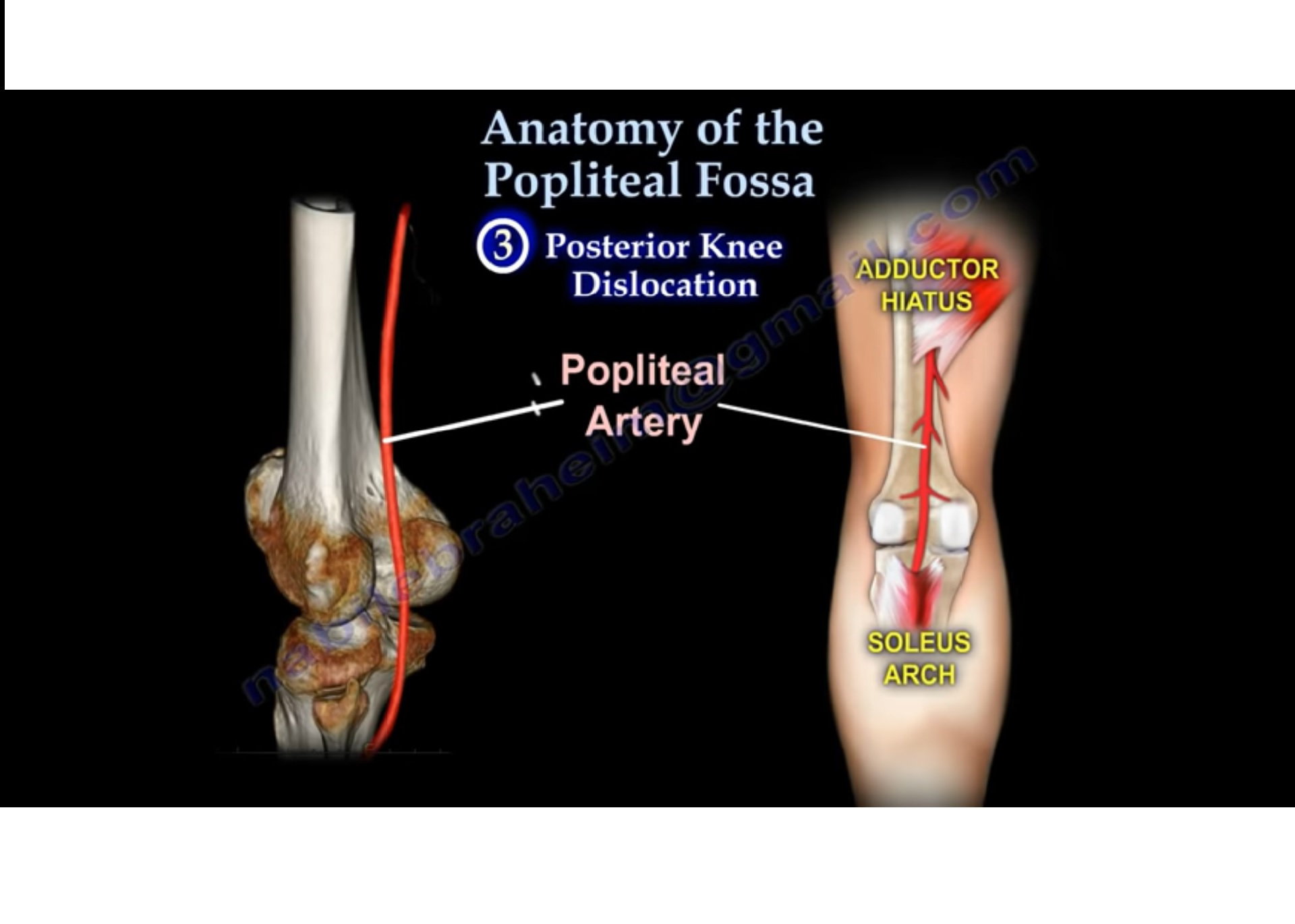
The fossa poplitea anatomy is home to several critical structures that are essential for the function of the lower limb. These include the popliteal artery, popliteal vein, tibial nerve, common fibular nerve, and lymphatics. Each of these components plays a unique role in maintaining circulation, sensation, and movement.
Popliteal Artery and Vein
The popliteal artery is a continuation of the femoral artery and is the primary blood vessel supplying the lower leg and foot. It branches into smaller arteries, such as the anterior and posterior tibial arteries, ensuring adequate blood flow to the muscles and tissues. The popliteal vein, located alongside the artery, facilitates the return of deoxygenated blood to the heart.
Tibial and Common Fibular Nerves
The tibial nerve and common fibular nerve are two major branches of the sciatic nerve that traverse the fossa poplitea anatomy. The tibial nerve provides motor innervation to the muscles of the posterior compartment of the leg and sensory innervation to the sole of the foot. The common fibular nerve, on the other hand, innervates the anterior and lateral compartments of the leg, contributing to dorsiflexion and eversion of the foot.
Together, these structures form a complex network within the fossa poplitea anatomy, ensuring the seamless operation of the lower limb. Their proximity to one another also makes the popliteal fossa a critical area for clinical evaluation and intervention.
Why is the Popliteal Fossa Important?
The fossa poplitea anatomy is not just a structural feature of the knee; it is a vital component of the body's functional mechanics. Its importance lies in its role as a conduit for nerves and blood vessels, as well as its involvement in movement and circulation. Understanding why the popliteal fossa is important can provide insights into its clinical relevance and practical applications.
What Role Does the Popliteal Fossa Play in Movement?
The popliteal fossa serves as a transition zone between the thigh and the leg, facilitating the transfer of forces during movement. The muscles surrounding the fossa, such as the gastrocnemius and hamstring muscles, work together to enable actions like walking, running, and jumping. The neurovascular structures within the fossa ensure that these muscles receive adequate blood supply and nerve signals to perform their functions.
How Does the Popliteal Fossa Support Circulation?
The popliteal artery and vein are central to the fossa poplitea anatomy, ensuring the efficient circulation of blood to and from the lower limb. The artery delivers oxygenated blood to the muscles and tissues, while the vein returns deoxygenated blood to the heart. This dual function is critical for maintaining homeostasis and supporting physical activity.
Common Conditions Affecting the Popliteal Fossa
The fossa poplitea anatomy is susceptible to a variety of conditions that can impact its function and the overall health of the lower limb. These conditions range from vascular issues like popliteal artery aneurysms to nerve-related problems such as entrapment syndromes. Understanding these conditions is essential for early diagnosis and effective treatment.
Popliteal Artery Aneurysms
A popliteal artery aneurysm is a localized dilation of the artery that can lead to complications such as thrombosis or embolism. This condition often requires surgical intervention to prevent further complications and restore normal blood flow.
Nerve Entrapment Syndromes
Nerve entrapment syndromes, such as tarsal tunnel syndrome, can occur when nerves within the fossa poplitea anatomy are compressed or irritated. Symptoms may include pain, numbness, or weakness in the affected limb. Treatment options range from conservative measures like physical therapy to surgical decompression.
How to Examine the Popliteal Fossa?
Examining the fossa poplitea anatomy is a critical skill for healthcare professionals. A thorough examination can help identify abnormalities, diagnose conditions, and guide treatment decisions. Here’s how to perform a comprehensive assessment of the popliteal fossa.
Palpation Techniques
Palpation is the primary method for examining the popliteal fossa. Begin by asking the patient to flex their knee slightly to relax the muscles. Use your fingers to feel for the popliteal artery pulse, which can provide insights into circulation. Check for any swelling, tenderness, or irregularities in the area.
Imaging Studies
In cases where palpation is inconclusive, imaging studies such as ultrasound or MRI may be necessary. These techniques can provide detailed views of the fossa poplitea anatomy, helping to identify conditions like aneurysms or nerve entrapments.
Fossa Poplitea Anatomy in Clinical Practice
The fossa poplitea anatomy is a cornerstone of clinical practice, particularly in fields like orthopedics, vascular surgery, and neurology. Its intricate structure and function make it a focal point for diagnosing and treating a wide range of conditions.
Applications in Surgery
Surgeons often rely on a detailed understanding of the fossa poplitea anatomy when performing procedures such as bypass grafts or nerve decompressions. Accurate knowledge of the anatomical landmarks and contents of the fossa is crucial for minimizing complications and ensuring successful outcomes.
Role in Rehabilitation
In rehabilitation, the fossa poplitea anatomy plays a key role in designing treatment plans for patients recovering from injuries or surgeries. Physical therapists use their knowledge of the fossa to develop exercises that target specific muscles and improve overall function.
Frequently Asked Questions About Fossa Poplitea Anatomy
What is the fossa poplitea anatomy? The fossa poplitea anatomy refers to the diamond-shaped depression at the back of the knee, housing critical neurovascular structures like the popliteal artery, vein, and nerves.
Why is the popliteal fossa important? The popliteal fossa is vital for movement and circulation, serving as a conduit for